All posts by lauraevann

Seagrass Microbiome Sampling

Recently the Seagrass Microbiome group has been wrapped up in sending (and receiving!) microbiome sampling kits. These kits are part of a larger collaborative project focused on re-sequencing of Zostera marina samples in conjunction with sequencing of additional marine and freshwater Alismatid species and their microbiomes. JGI recently sequenced and released the Zostera marina genome, and we are hoping to build on their efforts and explore population level variation within Zostera marina, as well as differences in genome content and structure between Zostera and other Alismatids, in conjunction with microbiome sequencing.

The sampling kits sent by the seagrass microbiome group have focused on the microbial aspect of this project. We have asked members of the Zostera Experimental Network (ZEN) as well as additional collaborators to sample both plant tissue for sequencing (coordinated through Jay Stachowicz and Jeanine Olsen) and microbiome samples. We are extremely excited about this sample set, as it covers populations of Zostera marina across many different environments, for which we already have extensive metadata through the ZEN group! We are requesting root, sediment (within the rhizosphere), and leaf tissue, as detailed in the diagram below (courtesy of Jeanine Olsen).
microbiome

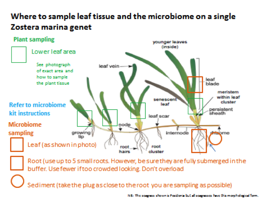
Collaborators are also sampling at two depths per site (deep and shallow), so that we can examine microbiome differences that may correlate with population depth. We are sampling 24 individuals per site, 12 per depth.

The kits are relatively straightforward and simple to both make and use, even if you’re not an experienced field microbiologist. We followed the kit and samplingl details we previously used (https://seagrassmicrobiome.org/protocols/microbial-sampling-kit/), with a few updates.

The kits now contain:
– 1 5cc syringe (for sediment collections)
– Tubes filled with Zymo buffer (DNA/RNA Shield)
– Plastic forceps
– Plastic spatula
– Parafilm
– Ethanol wipes

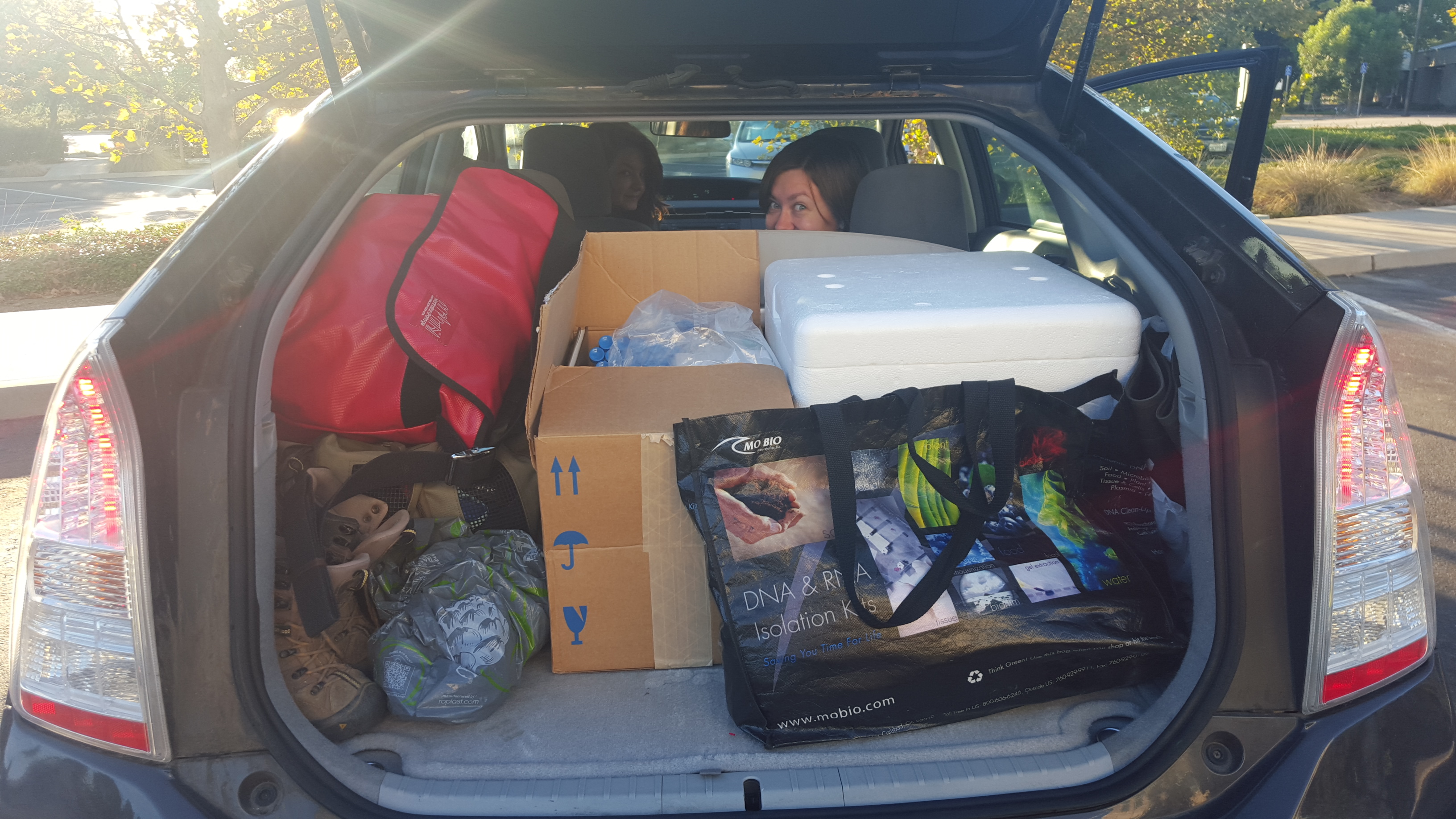
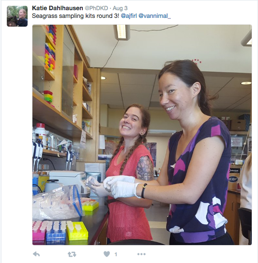
Here are a few photos of kit production:

Vann and Firl putting together kits in lab, photo from Katie Dahlhausen (@PhDKD)
Alana Firl and Laura Vann putting together kits in lab, photo from Katie Dahlhausen (@PhDKD)


Tubes all ready to go!


Close up of kit
Close up of kit


Completed kits, ready to go!

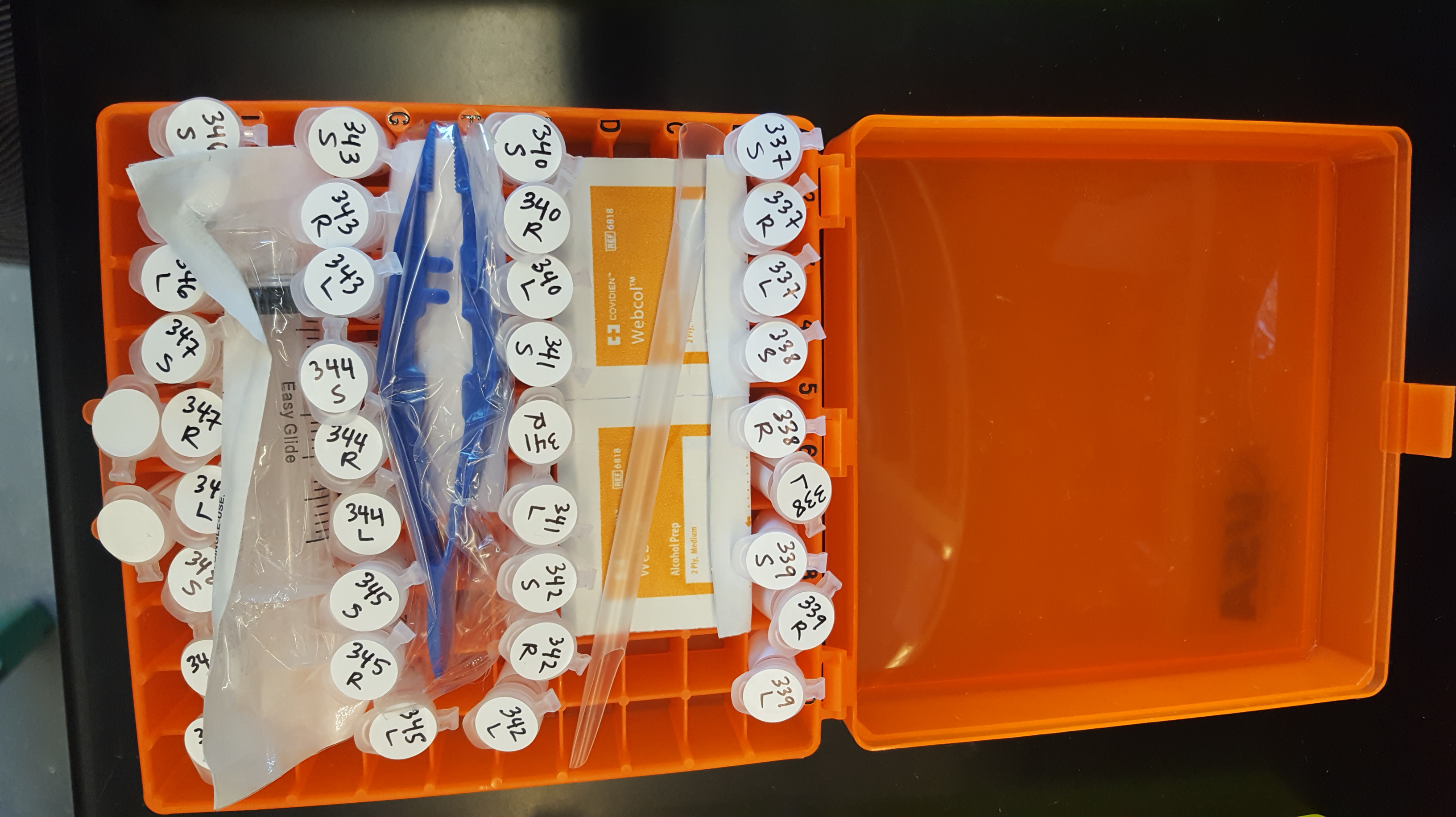
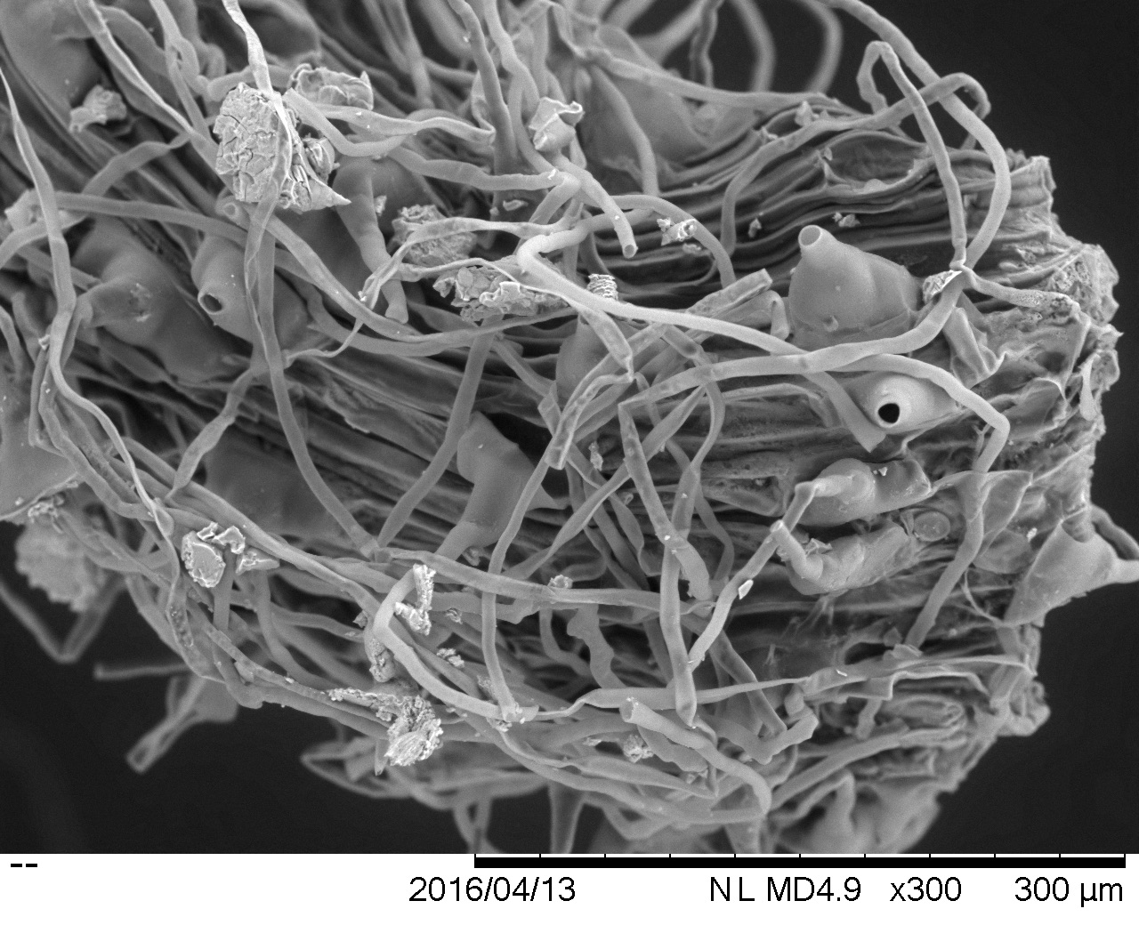
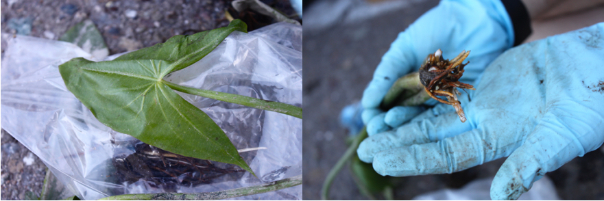
We have sent out all of the kits, and have already started receiving some completed samples in the mail. Here is a close up of some of the samples from Kotzebue, AK.

Samples for one individual from Kotzebue, AK. From left to right: root, sediment, and leaf.

A huge thanks to our collaborators for sampling, and to everyone from the Eisen lab who has helped make and send kits. Stay tuned for updates on sample processing and data !

Adventures in SEM

SEM demo room @ UCDavis. Hitachi TableTop is the large rectangular machine on the left.

Some of us at team Seagrass participated in a workshop on SEM (scanning electron microscopy) sponsored by the Electron Microscopy Lab at UC Davis. For this week only Hitachi Tabletop SEM was made available to researchers to test out. SEM is a great tool to produce high resolution images, especially for samples for which preparation techniques would otherwise alter the sample. We decided this would be a great opportunity to visually explore microbial diversity on seagrass roots/leaves/rhizomes. The amount of sample and preparation is minimal relative to other techniques. We’ve been particularly concerned with our previous FISH images, as FISH requires many washes, and may be removing microbes from the surface of our samples.

Samples on mount ready for SEM

We put a minimal amount of sample of root (top), fresh leaf (right), decaying leaf (bottom), and rhizome cross section (left) on a piece of carbon conductive paper (sticky on both sides), which was stuck on top of the SEM sample mount. We then inserted the mount into the SEM and turned on the vacuum. The vacuum for this particular SEM is actually a partial vacuum, which maintains a small amount of air molecules within the chamber, which produces a better image in the absence of coating your sample with conductors.

The results were great! We were able to see a lot of diatoms spread out across the leaf surface, as well as some interesting plaque (?) formations on the root tips. We also noticed differences in diatom abundance between the live and decaying leaves, with the live leaf being completely covered with diatoms. Diatoms are marine microbial eukaryotes (Heterokonts) that form silica based outer layers, often resembling complex geometric patterns. Diatom assemblages have previously been characterized in Thalassia testudinum and in Zostera marina.  We’re excited to use the SEM to explore microbial diversity on additional seagrass species and freshwater relatives within the Alismatales. Hopefully these results can help inform our future culturing experiments.

Root tip with strange filaments
Diatoms on rhizome surface
Close-up of diatoms on live leaf
Diatom distribution on live leaf
Diatom distribution on decaying leaf

CA sampling for project phylogeny

A few of us at Team Seagrass have been doing fieldwork in Northern CA in order to collect seagrass relatives. We have specifically been targeting freshwater species (highlighted in blue in the below phylogeny). We will continue with marine and brackish species when the tides in the San Francisco Bay are lower.

From https://phylogenomics.wordpress.com/people/post-docs/jenna-lang/seagrass/photo-4/: This is a phylogeny of the seagrasses and their aquatic relatives. This tree was built using parsimony and ~1200bp alignment of the rbcL gene. From Les and Cleland, 1997.
From https://phylogenomics.wordpress.com/people/post-docs/jenna-lang/seagrass/photo-4/: This is a phylogeny of the Alismatales, including the seagrasses and their aquatic relatives. This tree was built using parsimony and ~1200bp alignment of the rbcL gene. From Les and Cleland, 1997.

We have been following the methods detailed in https://seagrassmicrobiome.org/sample-collection-and-preservation/ and have managed to collect a few representative species from the following clades: PotamogetonaceaeAlismataceae, Hydrocharitaceae, Najadaceae, and the Lilaceae.

crap.001
Example leaf and root from Alisma species, collected at Cosumnes River

For further plant identification and the construction of a ‘host phylogeny’ we will be using chloroplast DNA markers.

In addition to what we have sampled thus far, we have also managed to sample some outgroups (MyriophyllumCeratophyllum, and Camboba). We will hopefully be able to sample additional outgroup species shown on the above tree.

We have been successful in 7 out of the 10 locations that we have tried. The lack of success so far can be attributed to drought conditions in CA and the increase in invasive submerged aquatic vegetation (e.g. Myriophyllum, Water Milfoil; and Eichhornia crassipes, Water Hyacinth). Water Milfoil and Water Hyacinth form dense mats below and above water, respectively, outcompeting native vegetation. Even in sites where we been successful, much of the area we surveyed has been overrun with these two invasive species.

Obviously drought has also been an issue for us, as many of the lakes and tributaries have little to no water in them this year, resulting in a lack of any aquatic vegetation. Water levels in Folsom Lake, for example, are so far reduced that the Park Service has had to build additional parking lots on the water side of the boat ramp in order for people to be able to get close to the new shoreline.

Map of Folsom Lake, with parking instructions drawn on by park official. X marks location of new parking lot, which is still at 15 minute walk to the shoreline.

In the following weeks we will head up to the foothills in search of remaining freshwater species, as well as explore the Bay Area coastline and salt marsh/Delta area for marine and brackish species. Stay tuned!

Sample preservation experiment

During ZEN DNA extractions we noticed that samples preserved in the Zymo buffer were forming a precipitate with the C1 solution from the MoBio kit. Furthermore, many of the samples also resulted in very low DNA yields, perhaps correlated with the precipitate formation. Any microbiologist will tell you that there are many different ways to preserve samples from the field, but there does not seem to be a universal *best* method.

We decided the best way to approach this problem for the Seagrass Microbiome project was to explore a variety of sample preservation methods and see which approximated the ‘real’ microbiome best (as measured by preservation on dry ice). We chose the following methods to try: Dry ice, Zymo, RNA-later, Drierite, and Ethanol. We also wanted to investigate how different preservation methods performed over time. We chose 4 time points post sampling for our extractions: 24 hours, 1 week, 2 weeks, and 1 month.

We drove to Putah Creek in Winters in search of submerged aquatic plants in the Alismatales (the order that contains the seagrasses). Here is a photo of our study site: 20150611_133052

We found a bed of Elodea canadensis growing near the shore and started sampling.

1460273_10205878523255831_7049527570566647813_n

We pulled out whole plants and divided them into root and leaf for each of our trial preservation methods, with 3 replicates per method per time point. We extracted DNA at each of the different time points, and did PCR of the 16S SSU rRNA gene, with subsequent library preparation and sequencing on the Illumina Mi-Seq. We used QIIME to analyze the sequence data and compare species diversity between our preservation methods.

PCA
Weighted unifrac PCoA

Our PCoA plot shows that preservation in Zymo best approximates the community captured using dry ice (our control), and that Drierite and Ethanol were both very different from anything else. RNA-later appears close to Zymo and dry ice, but we only have 5 data points for it. For the RNA-later and Ethanol extractions, we extracted directly from the solution without pelleting, which may have affected our end result.

Taxonomy summary plot: Order level

The taxonomy table supports the trend seen in our PCoA plot, with Drierite and Ethanol being very different, and Zymo, dry ice, and our 5 RNA-later time points showing similar communities.

Looking at variation in community structure across time points within Zymo we see little to no change.

Weighted unifrac PCoA showing time point variation within Zymo

However, we see some change in community structure by the month mark with dry ice.

Weighted unifrac PCoA showing time point variation within dry ice

Take home messages from our sample preservation experiment:

  • Using Drierite as a preservation technique does not capture the community assembly well.
  • RNA-later and Ethanol could have had better success with pelleting and removing the supernatant prior to extraction. We may investigate this further in the future.
  • In spite of our initial concerns regarding precipitate forming in the Zymo buffer, Zymo is the clear winner in our trial.
  • There are slight changes in the community assembly by the month mark, in all methods (depending on whether you use weighted or unweighted unifrac PCoA).
  • Due to the current ease of sampling using dry ice, we will continue this method except in situations where samples must be kept at room temperature (we will then use Zymo).

20150902_072734

HAPPY SAMPLING 🙂 Please see https://seagrassmicrobiome.org/sample-collection-and-preservation/ for more details regarding sampling and preservation protocols.